- Home
- Patient Care
- Services
- Shoulder & Elbow
- Overview
- Elbow Arthroscopy Information
- The Anatomy of the Elbow
The Anatomy of the Elbow
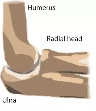
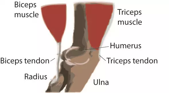
The elbow is a hinged joint made up of three bones, the humerus, ulna, and radius. The ends of the bones are covered with cartilage. Cartilage has a rubbery consistency that allows the joints to slide easily against one another and absorb shock. The bones are held together with ligaments that form the joint capsule. The joint capsule is a fluid filled sac that surrounds and lubricates the joint.
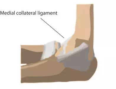
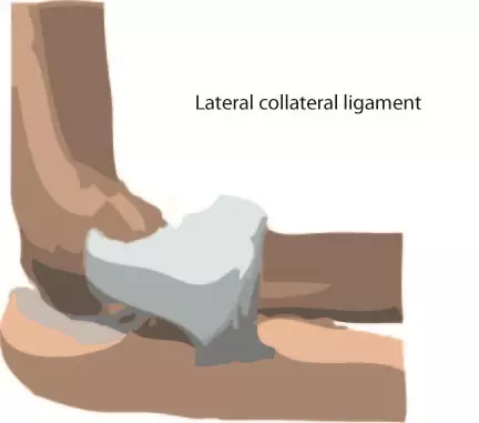
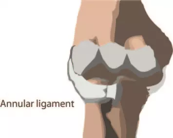
The important ligaments of the elbow are the medial collateral ligament (on the inside of the elbow) and the lateral collateral ligament (on the outside of the elbow.) Together these ligaments provide the main source of stability for the elbow, holding the humerus and the ulna tightly together. A third ligament, the annular ligament, holds the radial head tight against the ulna.
There are tendons in your elbow that attach muscle to bone. The important tendons of the elbow are the biceps tendon, which is attached the biceps muscle on the front of your arm, and the triceps tendon, which attaches the triceps muscle on the back of your arm.
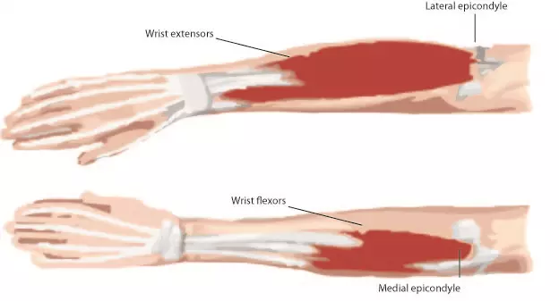
The muscles in your forearm cross the elbow and attach to the humerus. The outside (lateral) bump just above the elbow is called the lateral epicondyle. Most of the muscles that straighten the fingers and wrist come together and attach to the medial epicondyle, or the bump on the inside of your arm just above the elbow. These two tendons are important to understand because they are common locations of tendonitis.
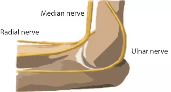
All of the nerves that travel down the arm pass across the elbow. Three main nerves begin together at the shoulder the radial nerve, the ulnar nerve and the medial nerve. These nerves are responsible for signaling your muscles to work and to also relay sensations such as touch, pain and temperature.
NEXT TOPIC: Common Conditions that Require Elbow Arthroscopy